We use epifluorescence predominantly to investigate the character and distribution of hydrocarbons trapped within fluid inclusions. Our system is equipped with a Zeiss HBO UV illuminator and “Epiplan Neofluar” objectives covering a wide range of magnifications (x1.25 up to x100). Using the digital camera attached to this microscope we are able to capture images of specific inclusions under the very low light levels that are commonly encountered during epifluorescence imaging.
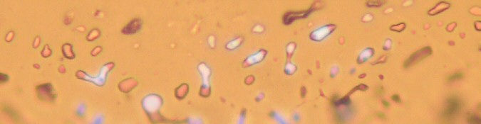
Image captured using mixed transmitted (plane polarised) light and incident UV (epifluorescence) illumination, showing a cluster of fluid inclusions trapped within vein calcite. Within this cluster, the inclusions trap different proportions of an aqueous fluid (non-fluorescent, so appears colourless in this image) and a brightly blue fluorescent hydrocarbon liquid. Where the inclusions only trapped the aqueous liquid, they have remained monophase at room temperature (i.e. have failed to nucleate a vapour bubble). In contrast, in the inclusions that trap a mixture of water and hydrocarbon, vapour bubbles are prominent within the hydrocarbon phase. (Field of view ~180um.)
The colour of the fluorescence is a reflection of the composition of the hydrocarbon, and can be used in a qualitative sense to approximate oil API gravity. Where samples have seen multiple generations of hydrocarbon of differing compositons / maturities, epifluorescence can be used to differentiate between different generations of fluids. This, combined with microthermometric analysis of the fluid inclusions, can be used to reconstruct filling / fluid histories.
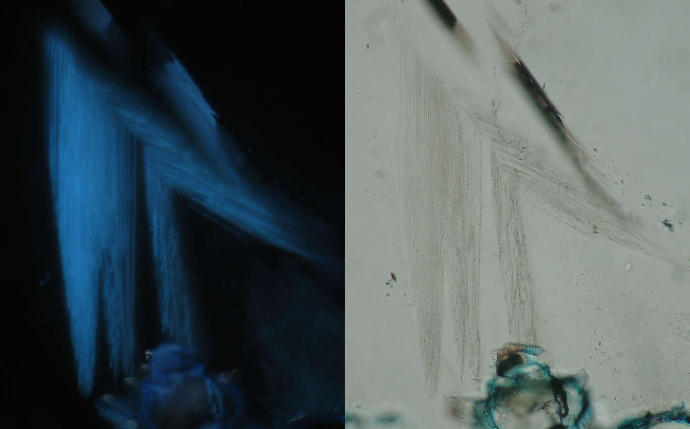
Paired epifluorescence and plane polarised light images showing planes of very fine, blue-fluorescent hydrocarbon inclusions trapped along growth planes in authigenic quartz (field of view of each image ~430um).
Sandstone petrography and reservoir quality specialists