 When we bought our bench-top SEM a few months ago, I thought it would be interesting to revisit this sample, which is one that I spent a lot of time looking at when I was a PhD student ( … back in the day … sigh). I also read a piece in today’s paper that gave me pause to think about just how much I (try not to) take for granted in my day-to-day work .
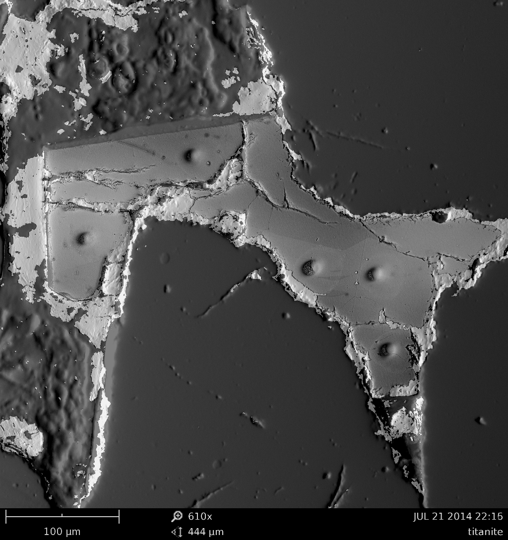
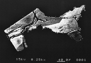
This sample is from the Cock of Arran and is from a Permian sandstone that is very unusual in that it contains the mineral titanite (A.KA. sphene) which precipitated as a pore-filling cement. (In the ~20 years I have been working in petrography since my PhD, I still have not encountered this mineral as a digenetic component in any other sandstone, in spite of looking really hard to find it.) This sample has had a pretty rough time of it. Following erosion, the sand grains were deposited in an arid environment during the Permian. They suffered burial diagenesis, near-contact metamorphism and deformation (due to the emplacement of the nearby Arran Granite), before being exhumed and exposed on a particularly wind-swept, midge-ridden coastline. This sample was collected from a beach exposure in 1994, and prepared as a polished thin-section by the lab at the University of Aberdeen. I then spent a couple of years subjecting it (and it’s contempories) to all manner of ignominies, under the watchful supervision of Malcolm Hole and Nigel Trewin.
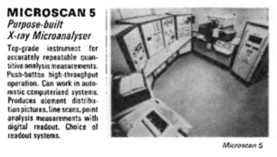
The analyses included optical and scanning electron microscopy and electron microprobe analysis (Cambridge Microscan 5; see above), as well as more advanced techniques such as radiogenic (Sm-Nd) isotope analysis (at SUERC), and ion microprobe analysis (using NERC / Edinburgh University’s Cameca ims-4f…another beast!).
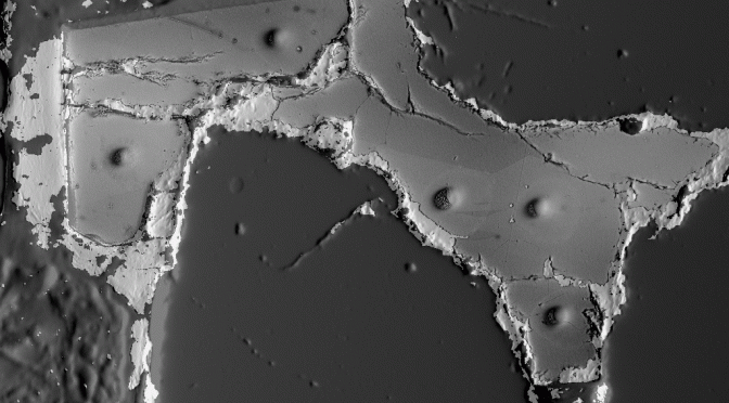
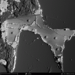
The new, featured, image is a composite derived from two images. One is a normal “compositional” BSEM image. The other is a “topographical” BSEM image. The image shows the sector zonation in the titanite, and also the amazingly precise holes drilled by the ion-probe. The bright material at the margins of the crystal, and trapped along some of the microfractures, is gold residua from the sputter coating applied in preparation for ion probe analysis (that I failed to remove during subsequent cleaning of the sample). The image also captures the irregularities in the sample surface caused by differences in hardness between the different minerals.